Intellectual Property & Publications
Intellectual Property (US Patent Applications)
1. Rahmat R, et al., Techniques for processing CBCT projections. US Patent App. 18/157,524 (2024).
2. Rahmat R, et al., Techniques for removing scatter from CBCT projections. US Patent App. 18/157,504 (2024).
3. Rahmat R, et al., Techniques for adaptive radiotherapy based on CBCT projection correction and reconstruction. US Patent App. 18/157,531 (2024).
Peer-Reviewed Publications
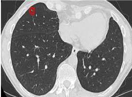
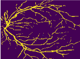
4. Rahmat R, et al., Radiomics-Led Monitoring of Non-small Cell Lung Cancer Patients During Radiotherapy. In 25th Annual Conference on Medical Image Understanding and Analysis 2021 (pp. 532-546). Springer.[Publisher's link] [Bibtex]
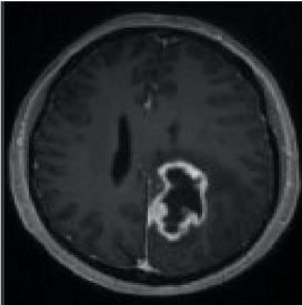
5. Wan Y, Rahmat R, Price SJ. Deep learning for glioblastoma segmentation using preoperative magnetic resonance imaging identifies volumetric features associated with survival. Acta Neurochirurgica. 2020 Jul 13:1-4.[Publisher's link] [Bibtex]
6. Rahmat R, Saednia K, Khani MR, Rahmati M, Jena R, Price SJ. Multi-scale segmentation in GBM treatment using diffusion tensor imaging. Computers in Biology and Medicine. 2020 May 22:103815.[Publisher's link] [Bibtex]
7. Stefani A, Rahmat R, Harris-Birtill D. Autofocus Net: Auto-focused 3D CNN for Brain Tumour Segmentation. InAnnual Conference on Medical Image Understanding and Analysis 2020 Jul 15 (pp. 43-55). Springer, Cham. [Publisher's link] [Bibtex]
8. Rahmat R, Brochu F, Li C, Sinha R, Price SJ, Jena R. Semi-automated construction of patient individualised clinical target volumes for radiotherapy treatment of glioblastoma utilising diffusion tensor decomposition maps. The British Journal of Radiology. 2020 Apr;93(1108):20190441.[Publisher's link] [Bibtex]
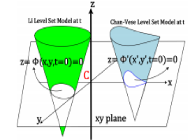
9. Rahmat R, Harris-Birtill D. Comparison of level set models in image segmentation. IET Image Processing. 2018 Aug 28;12(12):2212-21.[Publisher's link] [Bibtex]
10. Rahmat R, Nailon WH, Price A, Harris-Birtill D, McLaughlin S. New level set model in follow up radiotherapy image analysis. InAnnual Conference on Medical Image Understanding and Analysis 2017 Jul 11 (pp. 273-284). Springer, Cham.[Publisher's link] [Bibtex]
11. Rahmat R. Monitoring of lung cancer patients during radiotherapy using combined texture and level set analysis of CBCT images (Doctoral dissertation, Heriot-Watt University)[Publisher's link] [Bibtex]
12. Rahmat R, Yang F, William WH, McLaughlin S. Lung Tumour Segmentation using a Combined Texture and Level Set [Publisher's link]
13. Rahmat R, Malik AS, Kamel N, Nisar H. 3D shape from focus using LULU operators and discrete pulse transform in the presence of noise. Journal of visual communication and image representation. 2013 Apr 1;24(3):303-17.[Publisher's link] [Bibtex]
14. Rahmat R, Mallik AS, Kamel N, Choi TS, Hayes MH. 3D shape from focus using LULU operators. InInternational Conference on Advanced Concepts for Intelligent Vision Systems 2012 Sep 4 (pp. 237-245). Springer, Berlin, Heidelberg.[Publisher's link] [Bibtex]
15. Rahmat R, Malik AS, Faye I, Kamel N, Nisar H. An overview of LULU operators and discrete pulse transform for image analysis. The Imaging Science Journal. 2013 Feb 1;61(2):146-59.[Publisher's link] [Bibtex]
16. Rahmat R. Shape from Focus Using Lulu Operators and Discrete Pulse Transform in the Presence of Noise. [Publisher's link]
17. Rahmat R, Malik AS, Kamel N. 3-D content generation using optical passive reflective techniques. In2011 IEEE 15th International Symposium on Consumer Electronics (ISCE) 2011 Jun 14 (pp. 639-642). IEEE. [Publisher's link] [Bibtex]
18. Rahmat R, Malik AS, Kamel N. Comparison of LULU and median filter for image denoising. International Journal of Computer and Electrical Engineering. 2013 Dec 1;5(6):568.[Publisher's link] [Bibtex]
19. Rahmat R, Kamel NS, Yahya N. Principle Subspace-Based Signature Verification Technique using Reduced Sensors Data Glove. In2009 Innovative Technologies in Intelligent Systems and Industrial Applications 2009 Jul 25 (pp. 317-321). IEEE.[Publisher's link] [Bibtex]
20. Rahmat R, Kamel NS, Yahya N. Subspace-based signature verification technique using reduced-sensor data glove. In2009 IEEE Symposium on Industrial Electronics & Applications 2009 Oct 4 (Vol. 1, pp. 83-88). IEEE.[Publisher's link] [Bibtex]
21. Rahmat R, Online Signature Verification using SVD Method.[Publisher's link]